PERCULINE
endo joint
Endoscopic Visualization
Direct nerve view, precise RF energy delivery.Targeted Transection
Selective nerve destruction, lasting pain relief.Optimized Outcomes
Visually guided, enhanced treatment success.
Endoscopic Control
Selective Nerve Destruction
Enhanced Treatment Outcomes
Facet & SI Joint Denervation
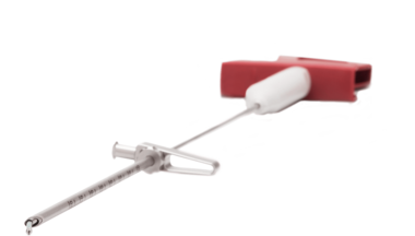
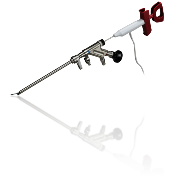
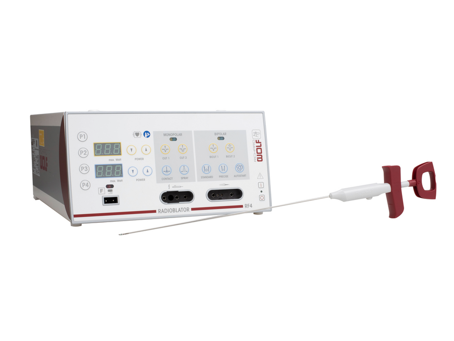
PERCULINE endo joint is specially developed instrument system for endoscopically controlled denervation of the facet and sacroiliac joint. In combination with the 4 MHz RADIOBLATOR RF generator, it offers a holistically convincing system solution. The visually controlled and precisely focused energy input also enables optimized treatment success. Endoscopically controlled denervation allows both the medial and lateral branches of the dorsal ramus to be visualized, safely transected and coagulated.
PERCULINE
Denervation of the facet and sacroiliac joint
Thermal denervation of the vertebral or sacroiliac joints involves interrupting the transmission of pain signals from the site of origin, i.e. the vertebral joints, to the brain by heating the pain fibers. In this way, local discomfort can be significantly reduced.
More information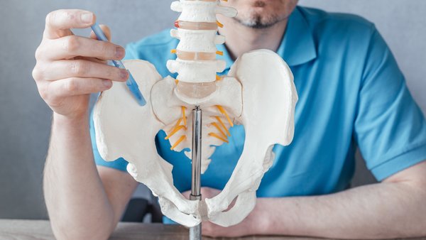
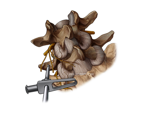
Denervation of the facet joint
The small facet joints are surrounded by a dense network of nerve fibers. These are irritated by inflammatory processes that occur, for example, in osteoarthritis.
Direct mechanical irritation can also occur due to instability. Leading symptoms are back pain or neck pain without significant radiation and without neurological deficits.
Pain is directed along the medial branches of the dorsal ramus spinal nerve. Therefore, the goal of 4 Mhz radiofrequency denervation is to selectively destroy these nerve fibers to permanently interrupt pain conduction.
Denervation of the sacroiliac joint
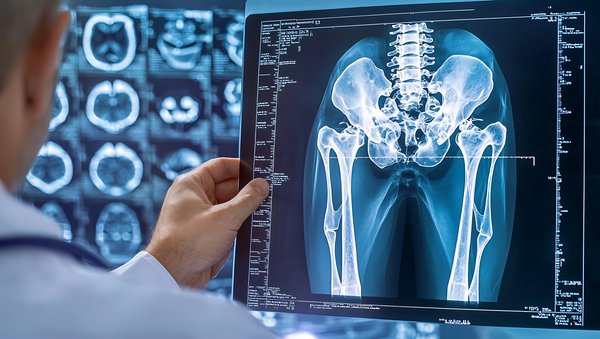
The sacroiliac joint is often a generator of back pain. Sacroiliac joint syndrome often occurs after spinal stabilization because of the increased stress placed on it after surgery. This in turn leads to mechanical and inflammatory irritation of the nerves at this joint.
Along the dorsal ramus of the spinal nerve, further transmission of pain is possible. These can be thermally eliminated at the point where the nerves leave the sacrum.
Treatment procedure
After local anesthesia, a puncture cannula is placed in the target area under X-ray guidance.
The puncture cannula is then replaced with a guide wire and a dilator is inserted over this guide wire.
After removal of the guide wire, the position of the dilator is checked under AP X-ray control and the working sleeve is inserted over the dilator.
The TipControl electrode and RF application are then inserted and denervation is performed via the foot switch. The procedure can easily be performed on an outpatient basis.
Note: This video contains medical images and scenes containing blood, which may be disturbing to some viewers.
Note acknowledgedHigh-quality Products
Special systems

Radiofrequency
surgical systems
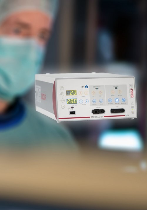
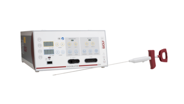
The RF4 radioblator with 4 MHz and TipControl enables precise, tissue-sparing coagulation.
More information